CERVICAL ARTERIAL DYSFUNCTION: TRANSFORMING THE PRACTICE TO ASSESS NECK PAIN

Cervical arterial dysfunction is an umbrella term that covers all known vascular pathologies and anatomical structures that may be compromised by assessment or management.
WHAT CAN HAPPEN TO BLOOD VESSELS?
Blood vessels can be subjected to:
- Inflammation
- Dissection
- Compression
- Stretch
- Narrowing
- Occlusion
- Aneurysm
- Vasospasm
- Atherosclerosis
- Endofibrosis
- Intimal damage
- Kinking
Dissection
During a vessel dissection, a tear occurs in the vessel wall where blood enters into the vessel wall. Separation of layers of the wall causes a false lumen where blood leaks into the vessel wall, forming the hematoma. It causes the lumen to become narrow, potentially leading to occlusion (1).

Reproduced from Kerry R, Taylor A. Haemodynamics
Dissection can occur to healthy blood vessels from trauma, usually blunt trauma.
DISEASED VESSEL
When a patient presents with a subjective history of previous cardiovascular or heart disease or taking statins or blood pressure medication, we may consider a diseased blood vessel with a risk of atherosclerosis, stenosis, thrombosis, embolus, ischemia, or aneurysm.
PATHOLOGIES UNDER THE UMBRELLA OF CERVICAL ARTERIAL DYSFUNCTION
Pathologies of all craniovertebral arteries come under cervical arterial dysfunction. The arteries that come under cervical artery dysfunction include (2):
- Carotid artery
- Vertebral artery
- Temporal artery
- Intracranial arteries/ circle of willis
THE PATHOLOGIES WE CAN CONSIDER IN CORONARY ARTERY DYSFUNCTION INCLUDE (2):
- Atherosclerosis
- Stenosis
- Aneurysm
- Distal emboli
- TIA
- Stoke
- Dissection
- Thrombus
- Distal emboli
- TIA
- Stroke
- Inflammation
- Arteritis
If you want to learn more about the signs and symptoms and presentation of individual cervical artery dysfunction, you can have a look at the lecture on Cervical Arterial Dysfunction by Alan Taylor on Trust-me Ed.
DIAGNOSTIC METHODS
- Subjective history
- Enhanced index of suspician
- Clinical examinaiton
- Duplex ultrasound
- Arteriogram
- MRA
- CTA
CLASSIFICATION OF CERVICAL ARTERIAL DYSFUNCTION
The cervical arterial dysfunction has been classified into five classes according to the Nottingham CAD classification model.

Reproduced from Kerry R, Taylor AJ. Cervical arterial dysfunction
IDENTIFYING THE CERVICAL ARTERIAL DYSFUNCTION (CAD) RISK FACTORS
We can identify the CAD risk factors from the patient’s history and physical examination.
History (identifying cad risk factors)
History of the patient can provide us with clues that the patient might have cervical arterial dysfunction. The majot risk factors include:
- Smoking
- Hypertesion
- Trauma
- Infection
- History of migraine
- Diabetes
- Anti-coagulation therapy
- Cardiovascular disease
If any of the above risk factors is present in the patient, consider the cervical arterial dysfunction a possibility.
Physical Examination
The physcal examination includes:
- Blood pressure measurement
- Cranial nerve assessment
- Eye examination
- Gait and proprioception assessment
- Balance assessment
Blood Pressure Measurement
Normal adult blood pressure is between 90/60mmHg and 120/80mmHg. Blood pressure is high if the blood pressure remains above 140/90mmHg consistently. A BP reading between 120/80mmHg and 140/90mmHg could mean the patient is at risk of developing high BP if the patient does not keep the blood pressure under control (3).
Why blood pressure measurement is important in Physical therapy patients?
Blood pressure measurement can help decide whether a patient is suited for a physiotherapy clinic or should be referred to the general physician or the emergency. In case of extremely high blood pressure, the patient might be at risk of stroke or cardiovascular accident (CVA), so he should be referred to the emergency.
The question arises whether Physical therapists should take blood pressure measurements. A study by Alan J. Taylor and Roger Kerry suggests that blood pressure measurement can be a great tool for risk factor assessment before manual therapy interventions (4).
A study has concluded that a 10 mm Hg reduction in systolic blood pressure reduces the risk of cardiovascular events by 20% (5).

What to do after blood pressure measurement?
NICE guidelines are in place to provide the care pathway for hypertensive patients (6). The figure below shows the blood pressure measurements and the respective path to deal with it.

Reproduced from the National Institute for health and care excellence (NICE)
The guidelines above show the pathway to diagnose and manage hypertensive patients. The pathway utilizes the triage method to separate the patient based on the severity of hypertension and directs the patients to their respective care facilities.
Cranial nerve examination
Cranial nerve examination is a significant part of cervical arterial dysfunction examination. 15-20% of vascular presentations will have cranial nerve involvement, especially in the later ischemic phase when we pick these signs and symptoms related to cranial nerve involvement.
Most clinicians argue that cranial nerve assessment takes time, but there are quick assessment techniques you can use to assess cranial nerves. So, cranial nerve assessment for cervical pain patients can be of significance.
Sensorimotor and Vestibular system examination
Sensorimotor and vestibular problems are more common than vascular problems in patients with traumatic neck pain. We can identify and manage these problems. Evidence suggests that sensorimotor dysfunction may lead to dizziness, visual disturbance, and balance impairments after trauma to the neck (7). If we can successfully identify these problems, we can manage them efficiently and improve the prognosis.
POST-TRAUMATIC NECK PAIN

Whiplash Associated Disorder and Traumatic Neck Pain
Whiplash injury patients present with a complex pattern. Studies show that our approach to managing whiplash and other neck injuries is not unsuccessful. In a study by Lamb et al, 2012, they provided ROM exercises, Manual therapy, soft tissue techniques, and advice to patients with a neck injury and pain, but the results were poor. The study concluded that a single session of advice from a physiotherapist appears beneficial in reinforcing the need to exercise and providing reassurance (8).
Psychological Issues in Patients with Traumatic Neck Pain
Patients with whiplash and other traumatic neck pain may present with various psychological problems, like general stress, fear of movement, anxiety, and post-traumatic stress disorder (PTSD). Assessing these may improve the prognosis.
Cold Hyperalgesia
Cold hyperalgesia in patients with traumatic neck pain suggests augmented central pain processing or neuropathic pain. It may be a predictor of poor prognosis (9).
Visual Disturbances
The whiplash-associated disorder may be associated with dizziness or visual disturbances linked to the disturbance of the sensorimotor system (10). Assessing these disturbances can make quite a difference in the prognosis.
Patient Presentation
Patients with whiplash or traumatic neck pain may present with:
- Dizziness
- Fear of movement
- Concern about pain and associated unusual features
- Complaint that their head feels heavy and they cannot lift their head off the pillow in the morning since the injury
MANAGEMENT OF NECK PAIN
The sooner we manage neck pain, the better outcome we get. If we delay the muscle activation of neck muscles after a neck injury, the muscles can accumulate fatty infiltrates and eventually become deactivated (11).

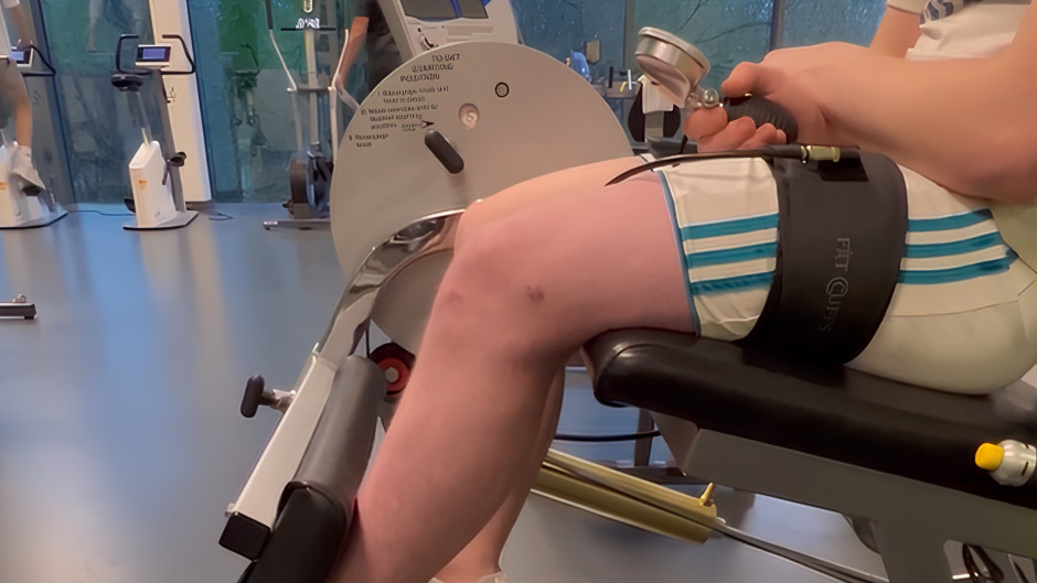
Strength Training
Strength training after neck injury improves isometric neck strength and reduces the risk of future injury (12). A review concluded that isometric neck training reduced match-related cervical spine injuries (13).
A study by Nazari G et al in 2018 concluded that deep and superficial flexor muscle training with home-based exercises is likely to reduce chronic neck pain and anxiety levels by a clinically relevant amount (14).
A study by Gaines and Cripps in 2017 concluded that neck muscle strengthening can reduce mild traumatic brain injuries (mTBIs) (15). Strengthening neck muscles can increase the response time in the cervical area and prevent further neck or brain injuries.
So, the strength training becomes an integral part of the neck pain rehabilitation. A boost in neck muscle isometric strength has significant potential to prevent subsequent neck injuries.
Sensorimotor Considerations
Visual-Vestibular Conflict
The sensorimotor problems in patients presenting with neck injury include visual-vestibular conflict. Normally, the visual and vestibular signals that govern the head movement are equal. But in neck injury or motion sickness, these signals may become unequal, resulting in dizziness, nausea, and vomiting.
The sensorimotor system works by compiling the information coming from different parts of the body. The conflict or mismatch of this information results in sensorimotor dysfunction, which can manifest as dizziness, balance impairment, and visual and sensory difficulties.
Major Vestibular Reflexes
Assessing the patient for major vestibular reflexes can help identify and cater to the problems effectively.
Three major vestibular reflexes are:
- Vestibulo-ocular reflex
- It keeps the eyes still while the head moves
- Vestibulo-colic reflex
- It keeps the head still or level while walking
- Vestibulo-spinal reflex
- It adjusts posture for rapid changes in position

Cervical Sensory Assessment
In patients presenting with neck pain, headache, and dizziness, assessing for the following can help prevent these acute symptoms from progressing to chronicity.
- Cervical spine – Joint position error (JPE)
- Oculomotor (gaze; eye movement)
- Postural stability
- Balance
Smooth pursuit eye movements
Smooth pursuit eye movements are the smooth movements of eyes when they follow a specific object. If these movements are not smooth abnormal smooth pursuit movements result, which indicates a possible ocular problem.
Smooth pursuit neck torsion
In smooth pursuit neck torsion, we observe the eye movements following a specific object with the neck in torsion. It gives us a better understanding of the combined working of the visual and vestibular systems.
REHABILITATION
We need a change in our current practice to better rehabilitate the patient. Use contemporary and updated research evidence in the practice. As discussed earlier in the blog post, vascular issues are rare in the case of neck pain.
Rehabilitating the patients sooner rather than later can prevent muscle deactivation and early return to sports or prior activity. Patients might have fear of movement and anxiety, and discussing these aspects of their problems with them can make the patient lose fear and get more involved in the rehabilitation process.
Integrating all aspects of the problem, including vestibular, ocular, psychological, balance, and strength into rehabilitation can make the rehabilitation program individualized and effective.
CONCLUSION
In conclusion, cervical arterial dysfunction is rare, but we should keep an index of suspicion. Assessing the patient thoroughly for sensorimotor and psychological issues can help us see the big picture of the problem and tackle it accordingly. Understanding the pathology and its mechanism becomes important during rehabilitation. Risk factor assessment can guide us toward the correct diagnosis. Integrating the research evidence with clinical practice can make our practice better and more effective. If you want to learn more about cervical arterial dysfunction and the management of patients with neck injuries, watch the lecture by Alan Taylor on Cervical Arterial Dysfunction on Trustme-Ed.
REFERENCES
- Robertson JJ, Koyfman A. Cervical Artery Dissections: A Review. J Emerg Med. 2016;51(5):508-18.
- Kerry R, Taylor AJ. Cervical arterial dysfunction: knowledge and reasoning for manual physical therapists. J Orthop Sports Phys Ther. 2009;39(5):378-87.
- Wales N. Blood pressure.
- Taylor AJ, Kerry R. Vascular profiling: should manual therapists take blood pressure? Man Ther. 2013;18(4):351-3.
- Ettehad D, Emdin CA, Kiran A, Anderson SG, Callender T, Emberson J, et al. Blood pressure lowering for prevention of cardiovascular disease and death: a systematic review and meta-analysis. Lancet. 2016;387(10022):957-67.
- Excellence NIfHaC. Hypertension in adults: diagnosis and management. 2023.
- Treleaven J. Dizziness, Unsteadiness, Visual Disturbances, and Sensorimotor Control in Traumatic Neck Pain. J Orthop Sports Phys Ther. 2017;47(7):492-502.
- Lamb SE, Williams MA, Williamson EM, Gates S, Withers EJ, Mt-Isa S, et al. Managing Injuries of the Neck Trial (MINT): a randomised controlled trial of treatments for whiplash injuries. Health Technol Assess. 2012;16(49):iii-iv, 1-141.
- Goldsmith R, Wright C, Bell SF, Rushton A. Cold hyperalgesia as a prognostic factor in whiplash associated disorders: a systematic review. Man Ther. 2012;17(5):402-10.
- Treleaven J, Jull G, Sterling M. Dizziness and unsteadiness following whiplash injury: characteristic features and relationship with cervical joint position error. J Rehabil Med. 2003;35(1):36-43.
- Elliott J, Pedler A, Kenardy J, Galloway G, Jull G, Sterling M. The temporal development of fatty infiltrates in the neck muscles following whiplash injury: an association with pain and posttraumatic stress. PLoS One. 2011;6(6):e21194.
- Geary K, Green BS, Delahunt E. Effects of neck strength training on isometric neck strength in rugby union players. Clin J Sport Med. 2014;24(6):502-8.
- Hrysomallis C. Neck Muscular Strength, Training, Performance and Sport Injury Risk: A Review. Sports Med. 2016;46(8):1111-24.
- Nazari G, Bobos P, Billis E, MacDermid JC. Cervical flexor muscle training reduces pain, anxiety, and depression levels in patients with chronic neck pain by a clinically important amount: A prospective cohort study. Physiother Res Int. 2018;23(3):e1712.
- Gaines A, Cripps A. Effectiveness of Neck Strengthening Exercises on Reducing Brain Injury. Journal of Sports Medicine and Allied Health Sciences: Official Journal of the Ohio Athletic Trainers Association. 2017;3(1).